Endocavitary After UAE for the Uterine Fibroid
Authors:
Byung Chul Kang
Clinical History:

Axial and sagittal MPR CT images (a and b) show a large defect in the posterior wall of infrarenal IVC with surrounding hematoma containing pseudoaneurysm (asterisks). There is a fistulous communication (arrow) between ruptured left second lumbar artery on volume rendering image (c)


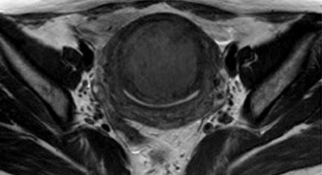
1A. T2 TSE TRA : T2-weighted MR image shows a huge mass of low signal intensity. B. T2 FS CORONAL C. T2 TSE SAGITAL


A & B. Uterine artery angiography shows bilateral hypertrophied uterine artery. C. After uterine artery embolization


Hypertrophied uterine artery could not be seen.


Submucosal fibroids with an interface dimension ratio on pre-procedural MRI of greater than 0.55 are more likely to migrate into the endometrial cavity. The majority of these do not cause significant additional symptoms. But, larger submucosal fibroids greater than 6 cm that become endocavitary may occasionally lead to post-procedural problems requiring further intervention and medical treatment.

- + Figure 1
-
Axial and sagittal MPR CT images (a and b) show a large defect in the posterior wall of infrarenal IVC with surrounding hematoma containing pseudoaneurysm (asterisks). There is a fistulous communication (arrow) between ruptured left second lumbar artery on volume rendering image (c)
- + Figure 2
-
1A. T2 TSE TRA : T2-weighted MR image shows a huge mass of low signal intensity. B. T2 FS CORONAL C. T2 TSE SAGITAL
- + Figure 3
-
A & B. Uterine artery angiography shows bilateral hypertrophied uterine artery. C. After uterine artery embolization
- + Figure 4
-
Hypertrophied uterine artery could not be seen.
- + Figure 5
-
Submucosal fibroids with an interface dimension ratio on pre-procedural MRI of greater than 0.55 are more likely to migrate into the endometrial cavity. The majority of these do not cause significant additional symptoms. But, larger submucosal fibroids greater than 6 cm that become endocavitary may occasionally lead to post-procedural problems requiring further intervention and medical treatment.